

Hearing breakthrough comes through studying mouse inner ear
Keck School of Medicine of the University of Southern California
Los Angeles, Calif.
Researchers at the Keck School of Medicine of the University of Southern California, in collaboration with Baylor College of Medicine, made a major discovery in understanding how the brain may help regulate hearing. And they did it by adapting an imaging technique currently used in ophthalmology offices, called optical coherence tomography.
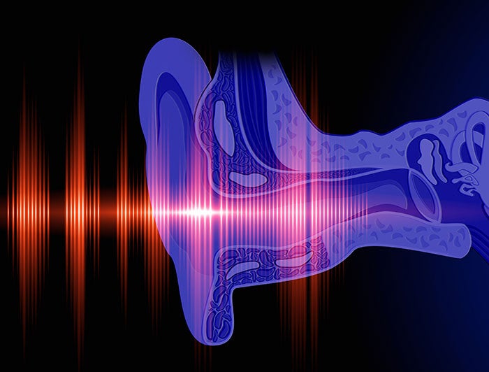
The team used OCT to capture real-time images of the cochlea — a part of the inner ear involved in hearing — in mice, revealing that the brain can send signals to the inner ear to enhance sound sensitivity. The study found that while the cochlea doesn’t respond to short-term brain state changes, it does increase activity in response to long-term hearing damage in mice with genetic hearing loss. The study suggests the brain compensates for hearing loss by boosting the function of remaining sensory cells.
The team is now preparing clinical trials to test whether blocking certain brain-to-ear signals could help reduce symptoms in patients with sound sensitivity disorders. Findings could lead to new treatments for conditions like tinnitus (ringing, buzzing or other phantom sounds) and hyperacusis (where everyday sounds become uncomfortably loud).